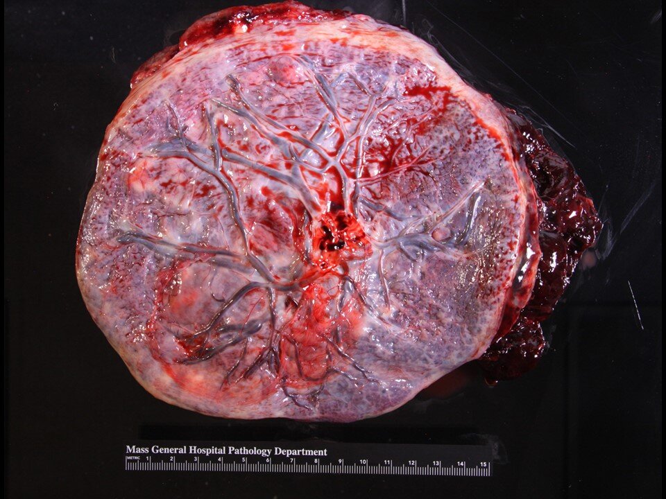
Gross placenta - Basic elements - Umbilical cord, membrane, placental disc
Accessory lobe, with transgressing vessels
Accessory lobe, with transgressing vessels
Accessory lobe, with transgressing vessels. Umbilical cord with velamentous / membranous insertion and thrombus in a velamentous vessel.
Bilobed placenta. Velamentous / membranous insertion of the umbilical cord. Note also the yolk sac remnant (circled).
Marginal umbilical cord insertion (defined as <1cm from disc edge)
Velamentous / membranous insertion of umbilical cord.
Marginal and membranous insertion of furcate umbilical cord. Furcate means that the umbilical cord vessels diverge prior to insertion.
Marginal and membranous insertion of furcate umbilical cord.
Furcate umbilical cord. Note that some of the vessels seen on the fetal surface appear poorly perfused.
Furcate umbilical cord. A probe illustrates this point. Specimen has been fixed in foramlin.
Hypocoiled umbilical cord with abnormal separation of the umbilical arteries – one of the umbilical arteries is separated from the bulk of Wharton’s jelly and is attached to the main cord by a thinned region
Hypercoiled umbilical cord.
Umbilical cord with yellow-green discoloration - meconium.
False knots - this finding has no clinical significance.
True knot - this finding may or may not be clinically significant, depending on several factors including how tight the knot is.
Edematous umbilical cord
Umbilical cord with false knots. Placenta with circummarginate membrane insertion - the membranes insert inward from the margin of the placental edge. This finding has no clinical significance.
Partially circummarginate (~70%)
Circumvallate membrane insertion - this finding is clinically significant. Like circummarginate membrane insertion, the insertion is inside the edge of the disc. However, there is a firm ridge at the site of insertion. It is associated with marginal hemorrhage / bleeding.
Circumvallate membrane insertion
Circumvallate membrane insertion - A cut section illustrates the point that circumvallate membrane insertion is characterized by a raised ridge.
Amniotic web / sail
Chorionic cysts
Chorionic cysts
Vernix caseosa / squamous metaplasia
Squamous metaplasia. Also note the membranes are dull, mucoid, green, and thickened and the umbilical cord has a yellow-green tinge, consistent with meconium.
Chorangioma - Cut section demonstrating a nodule with a red / spongy appearance.
Fetal papyraceous - remains of a demised twin that is retained in-utero after intrauterine fetal demise.
Normal maternal surface - will normally have some blood clot material that is relatively easily removed and does not cause indentation of the placental parenchyma (as opposed to true hemorrhage / abruption). Note the well formed cotyledons.
Disrupted maternal surface - the cotyledons are disrupted here and fragmented. It is important to note disruption as there is a chance for retained placenta.
Maternal surface with adherent blood blot. The histology showed features consistent with the clinical diagnosis of abruption, which included villous compression and both intervillous and intravillous hemorrhage.
Increased fibrin deposition seen on the maternal surface here. Histologically, consistent with massive perivillous fibrin deposition (also see below)
Increased perivillous fibrin deposition on cut sections - Massive perivillous fibrin deposition.
Infarction hematoma
Intervillous thrombi and infarction hematomas
Intervillous thrombi - some can be laminated
Multiple subchorionic plaques / subchorionic thrombi


