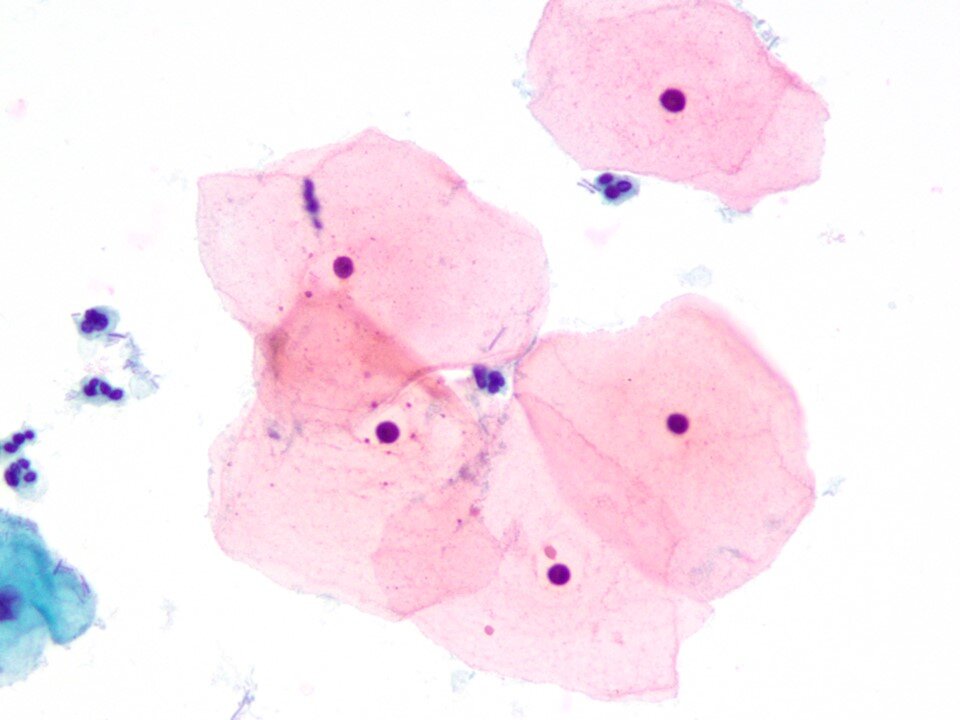
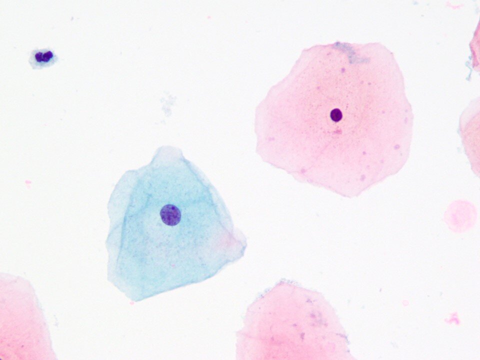
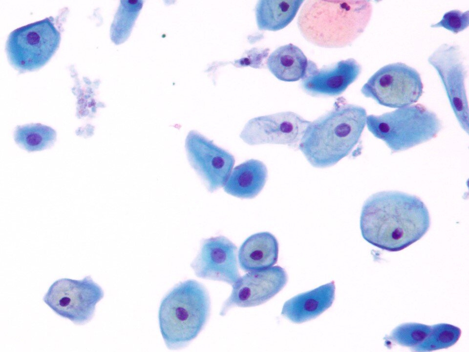
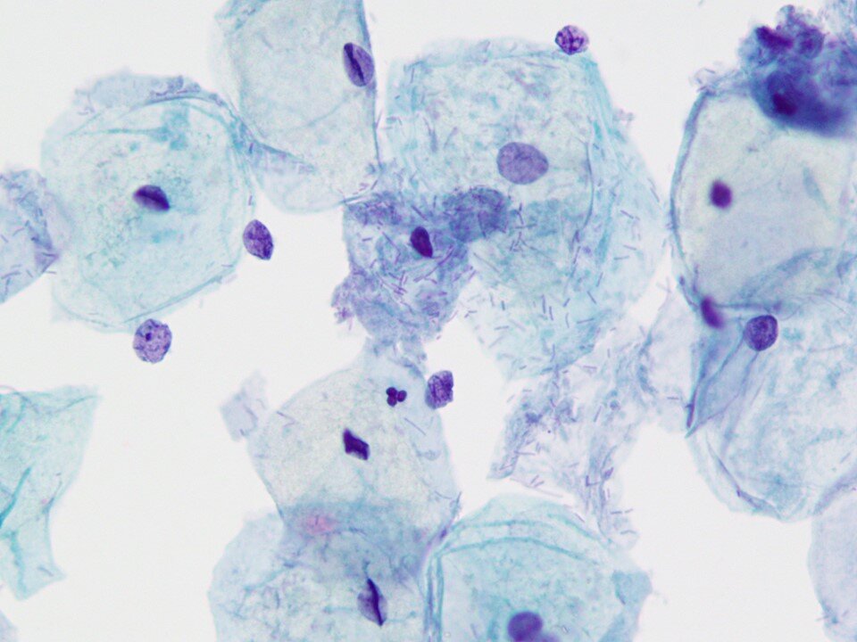
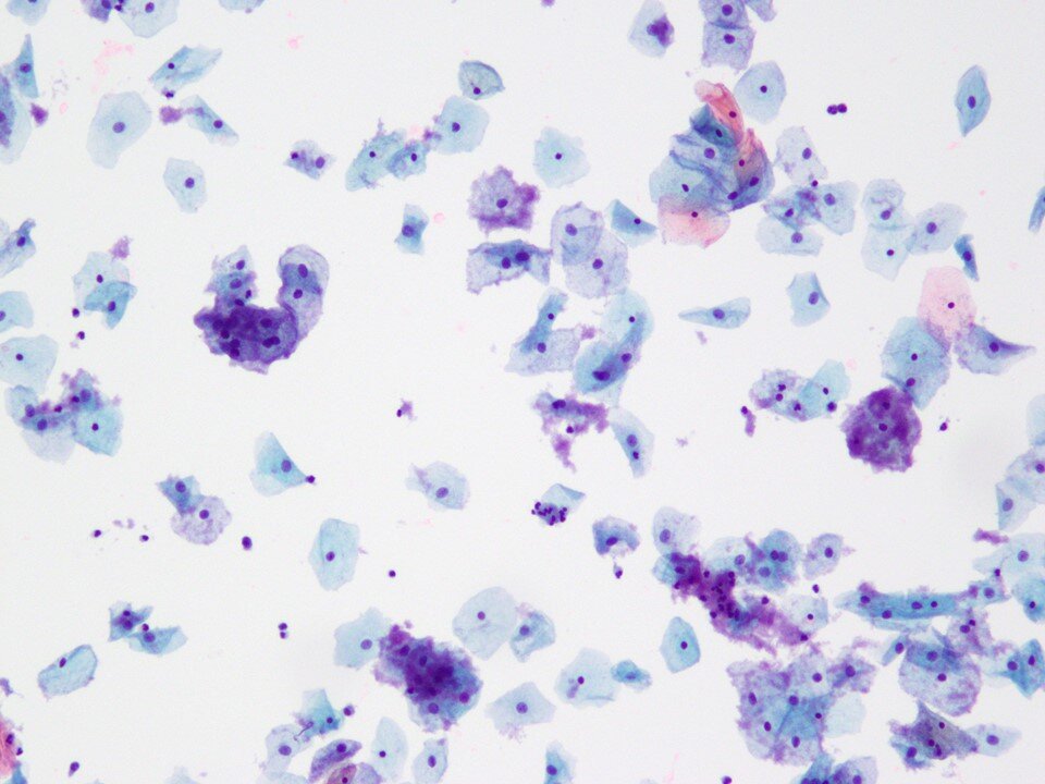
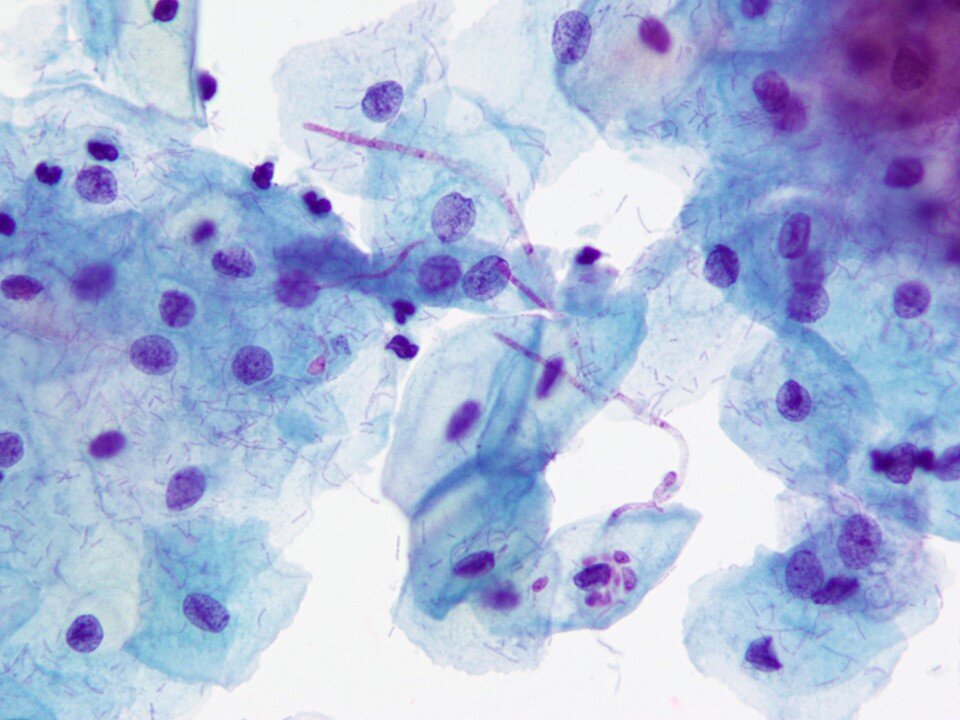
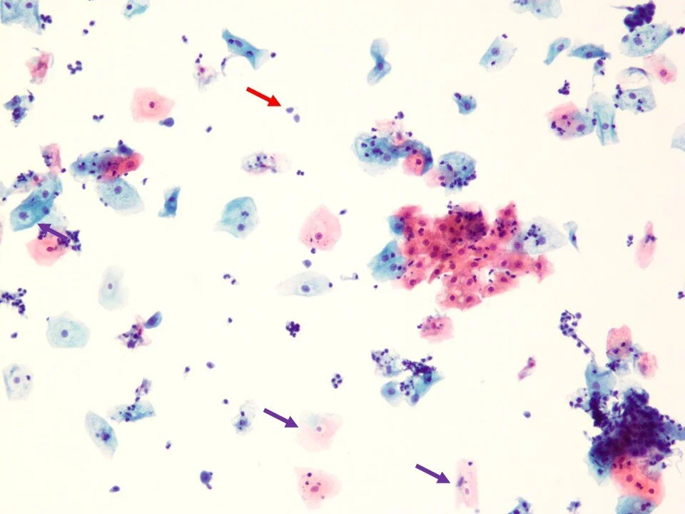
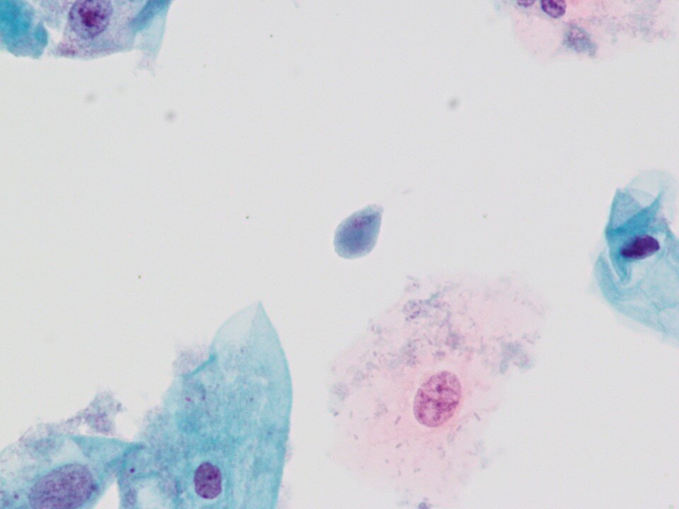
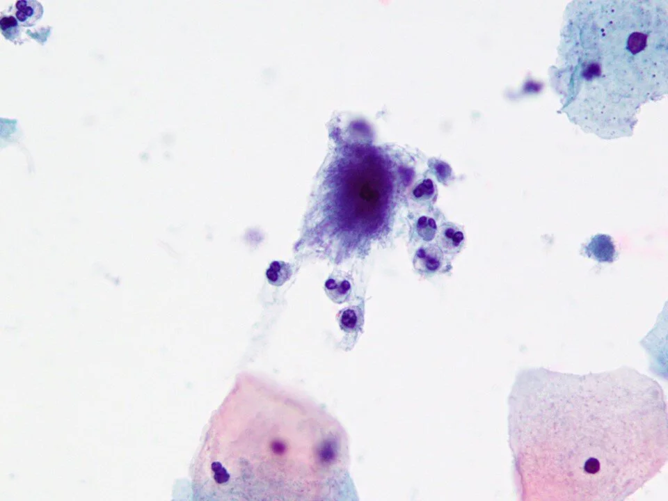
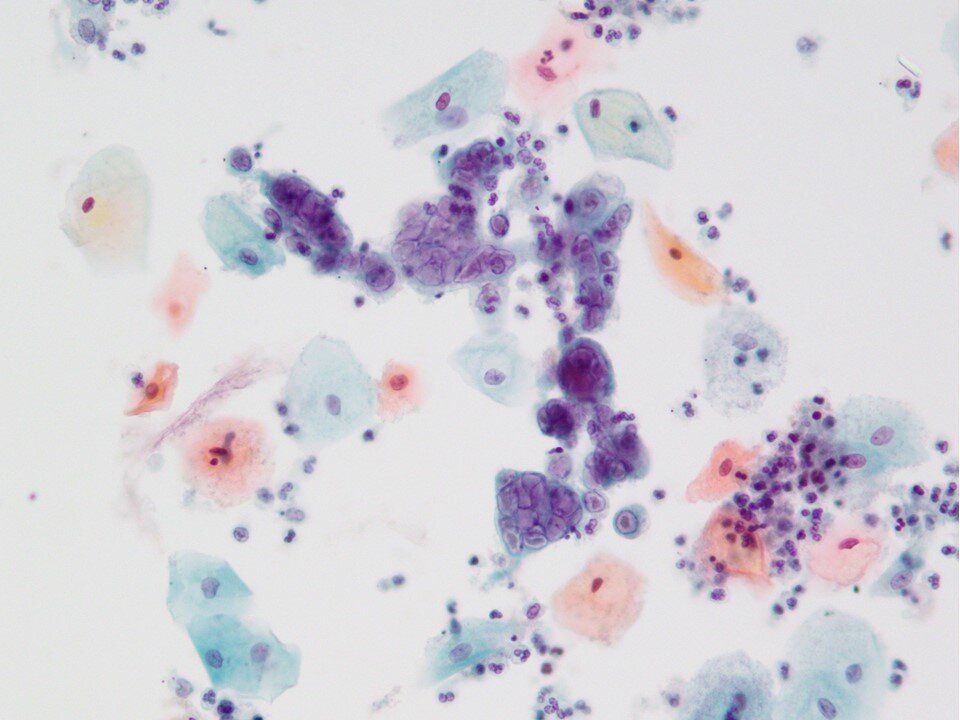
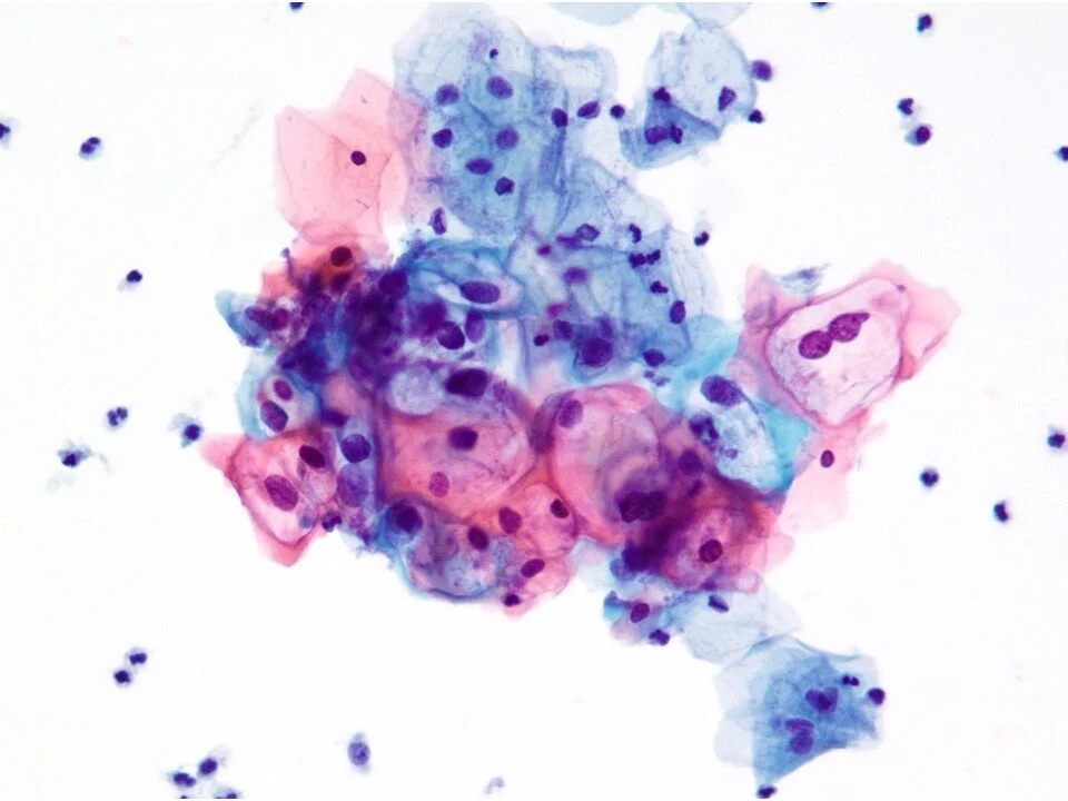
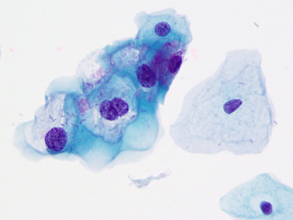
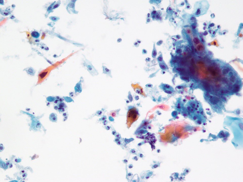
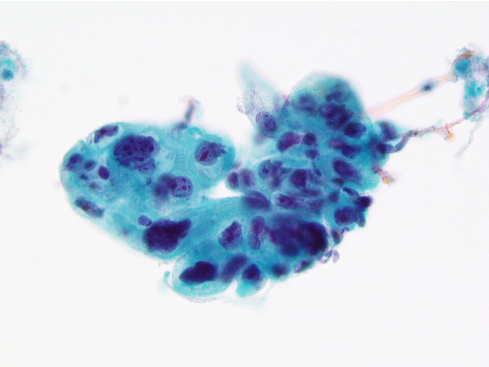
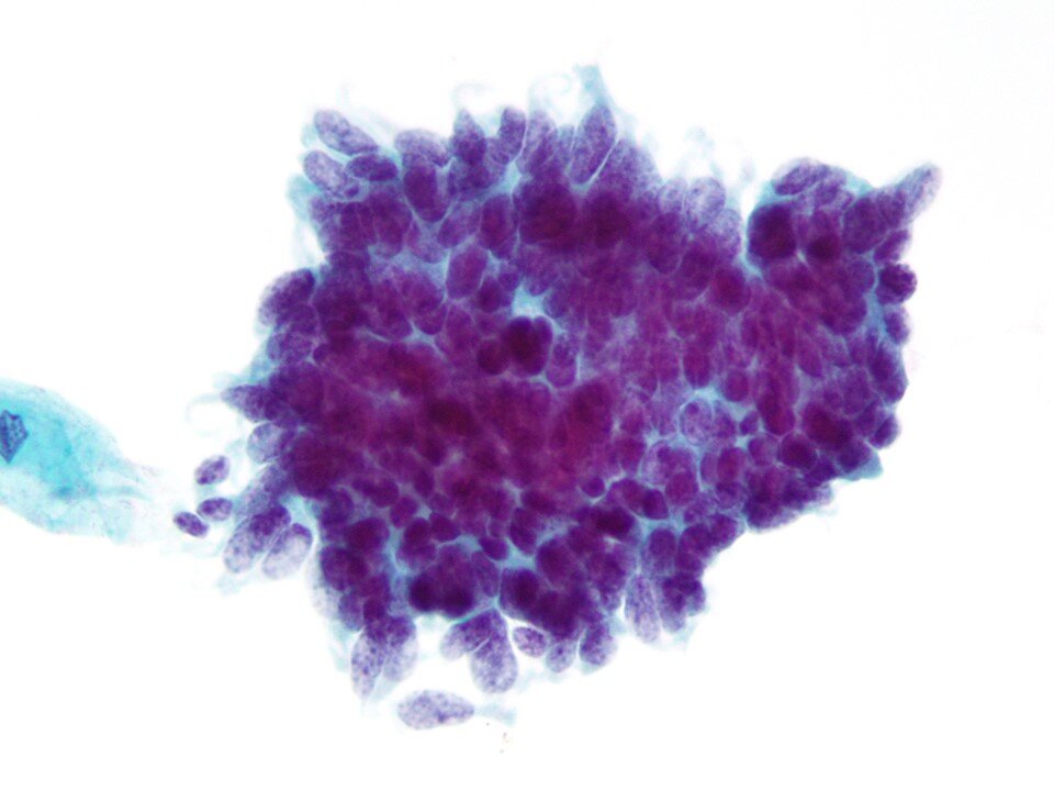
Superficial squamous cells - Large polygonal cells with transparent pink or blue/green cytoplasm and small pyknotic nucleus 5-6 microns in diameter. A predominant cell in samples from women of reproductive age.
Intermediate squamous cell - Large polygonal cell with transparent pink or blue/green cytoplasm with larger nucleus 8 microns in diameter that is not pyknotic. Occasionally binucleated or even multinucleated. A predominant cell in samples from women of reproductive age.
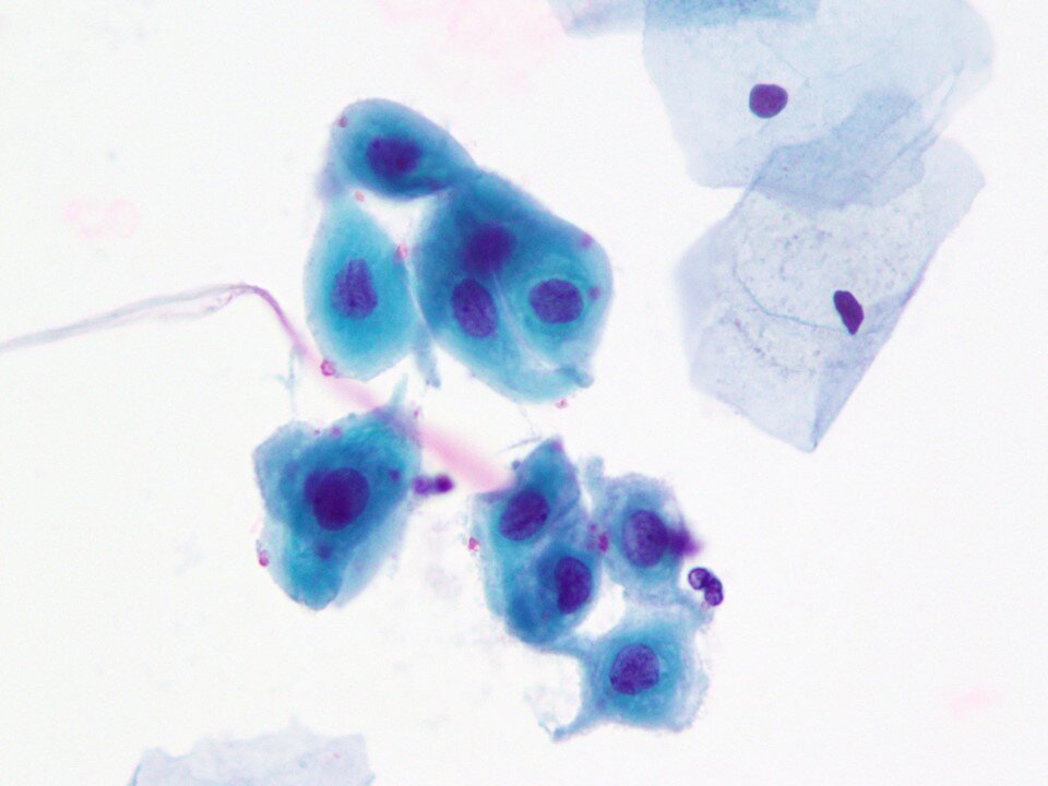
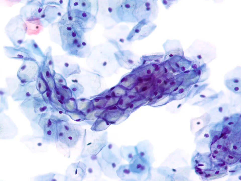
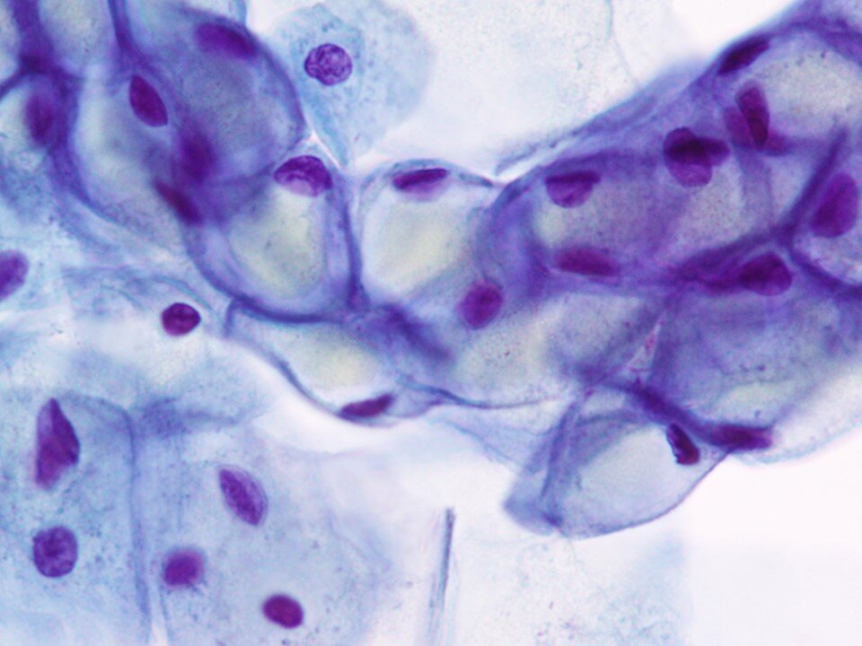
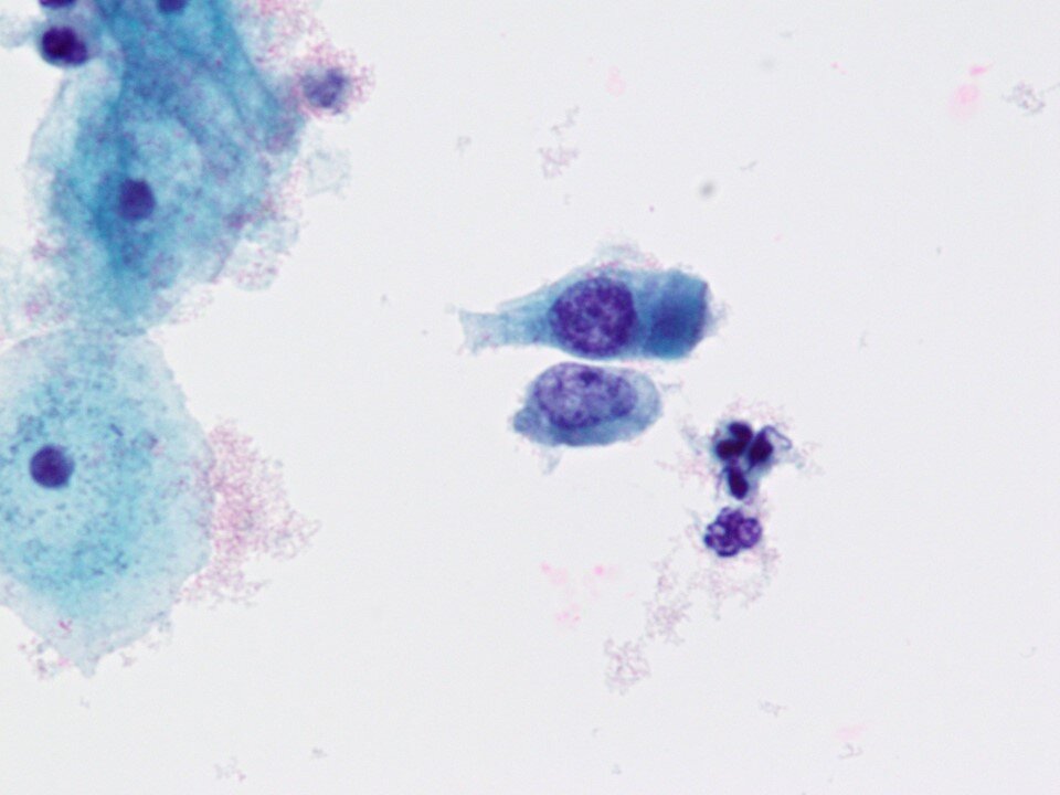
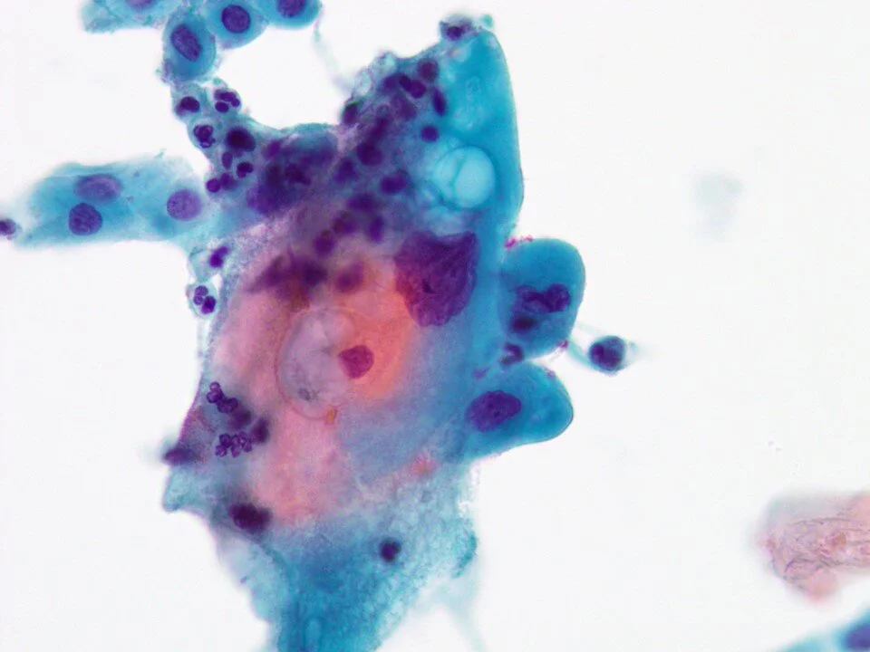
Squamous metaplastic cells - Common morphologic alteration of the endocervical epithelium, usually limited to the transformation zone. Squamous metaplastic cells have a characteristic aqua cytoplasm. They may show mild variation in nuclear size, slightly irregular nuclear contours, and slight hyperchromasia. They can be found singly or arranged as flat sheets in an interlocking fashion like paving stones.
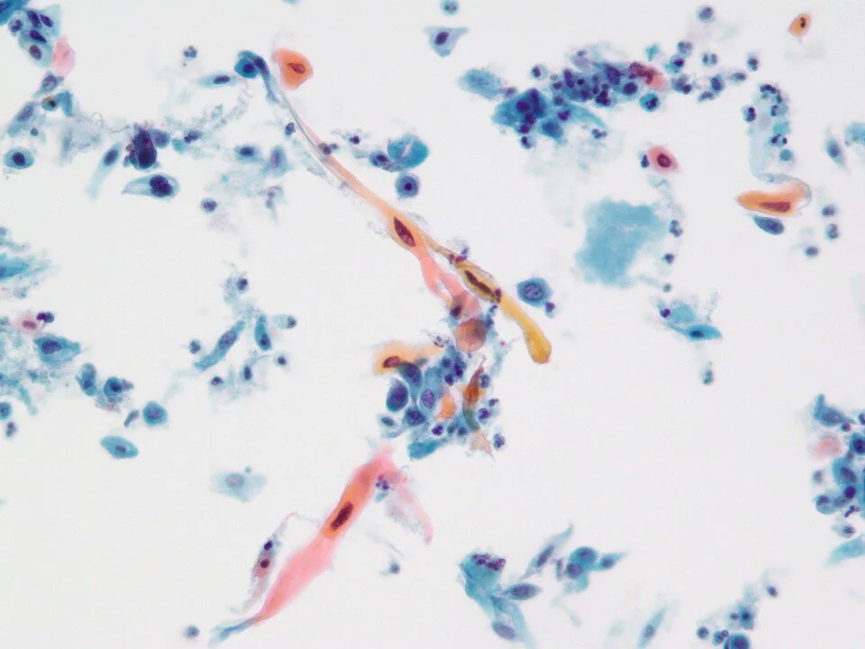
Squamous metaplastic cells - Cells having spindled cytoplasmic projections (“spider cells”) can be seen.
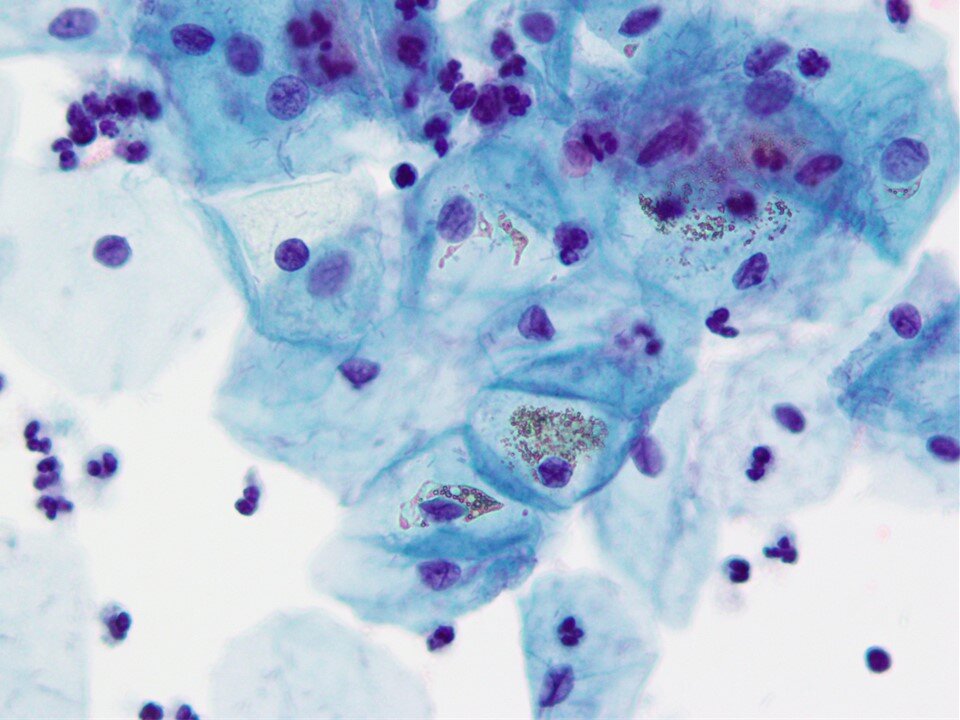
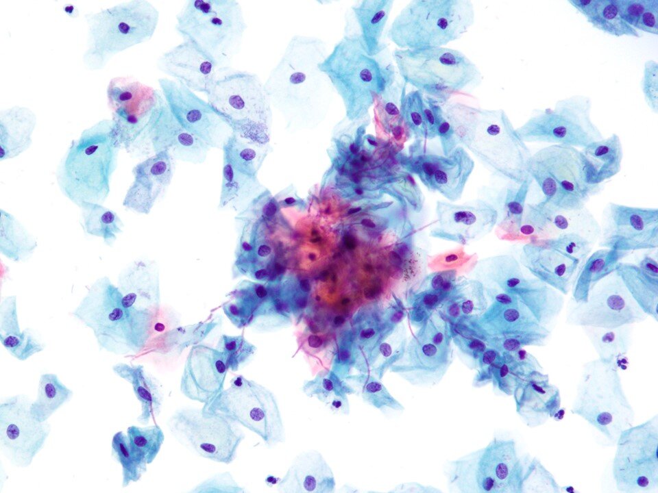
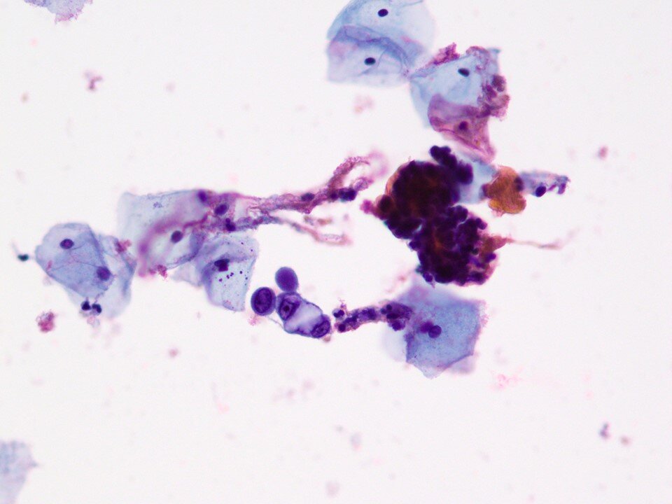
“Cornflaking” - A refractile brown artifact that results from bubbles of trapped air. It can be reversed by reprocessing/restaining the slide.
Parabasal cells - Immature squamous cells.
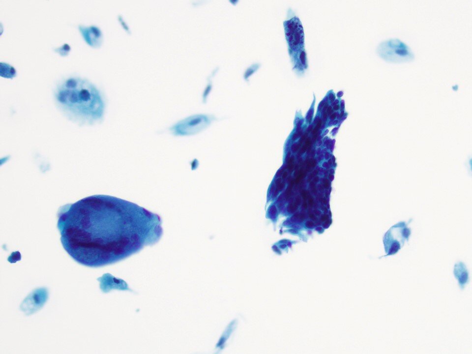
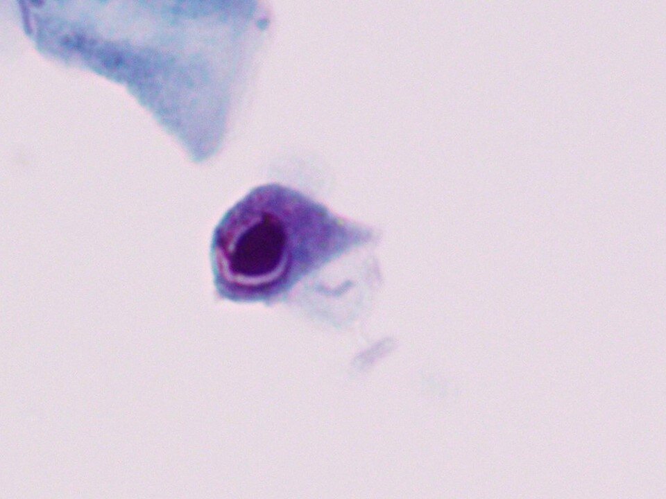
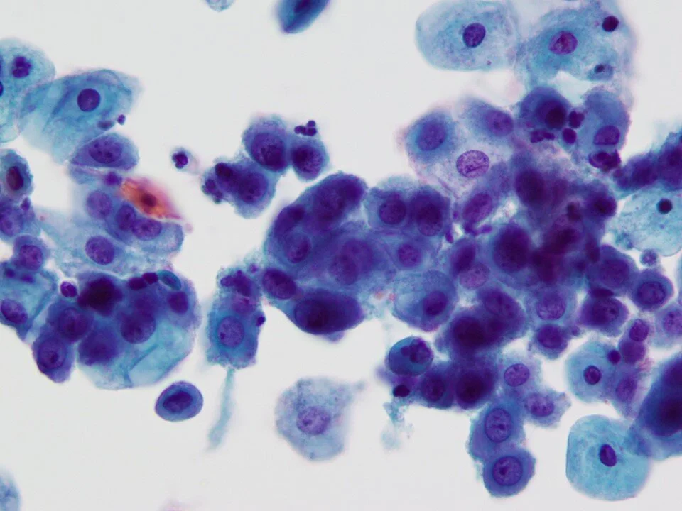
Basal cells - Immature squamous cells - a sign of atrophy (seen here in a hyperchromatic group). Encountered in low estrogen states such as premenarche, pospartum, postmenopause (as in this case), Turner Syndrome, status post bilateral oophorectomy, and administration of exogenous (androgeneric) hormones. The cellular components of atrophy, in addition to parabasal and basal cells, includes histiocytes / multinucleated giant cells and granular debris (not to be confused with the necrotic debris more typical of malignant processes).
Atrophy - Hallmark cells are basal and parabasal cells. Can be found as isolated cells or in hyperchromatic groups (as seen above). Some features (such as high N:C) may overlap with atypical processes such as HSIL. However, some features that help point to a benign process include finely granular and evenly distributed chromatin texture, smooth nuclear contours, and the absence of significant mitotic activity.
Navicular cells - intermediate cells with a “boat” shape. Associated with pregnancy or androgenic atrophy due to progesterone in women. Important to beware of as may mimic LSIL.
Endocervical cells - flat sheet.
Endocervical cells - “picket fence”
Endometrial cells - tight clusters are the typical appearance. Endometrial cells may be present in pap smears due to a variety of processes, including menstrual shedding in young women. They should be noted when identified in pap smears from women above the age of 45. In this age population, they may still be associated with benign processes and other processes such as polyps, but they have a higher likelihood of being associated with neoplastic / malignant processes (at least compared to younger women). The age cutoff of 45 was chosen as one that is most specific and sensitive based on available studies.
Endometrial cells - Nuclei that appear to be wrapping around at the periphery of the cluster are helpful clues, although it is a nonspecific finding.
Tubal metaplastic cells - In tubal metaplasia, the endocervical epithelium is replaced by an epithelium that recapitulates fallopian tube epithelium. The cells are columnar in shape and a characteristic feature is the presence of apical terminal bars and cilia.
